
OCAD University
“Making Digital Matter”

The General Idea
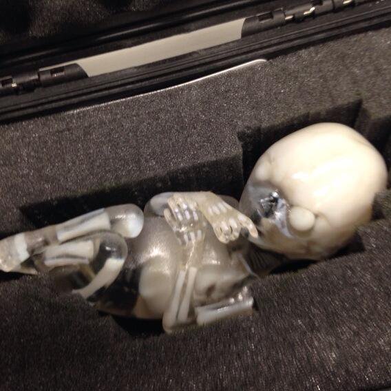
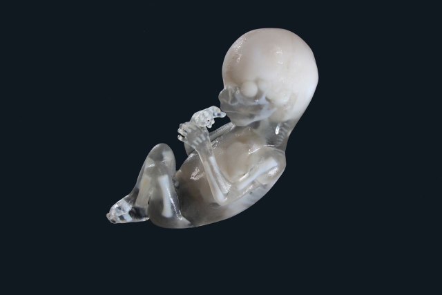
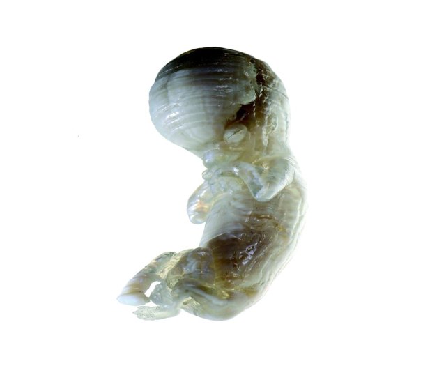
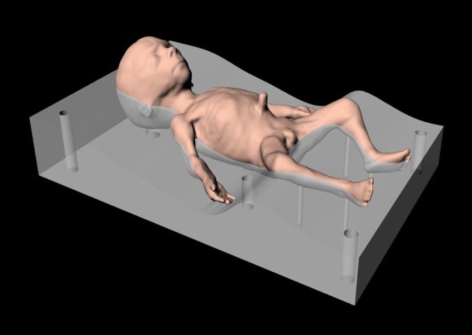
Medical surgeons need to hone their skills prior to performing high-risk surgical procedures on fetuses in utero and on newborns. To date, highly representational 3-dimensional fetal models, that have life-like physical properties, do not exist. The goal of our research project is to create a series of accurate and responsive fetal models to help surgeons visualize the complex physiology of a newly developing fetus and their associated pathologies. These models will provide surgeons with the visual and tactile information necessary to confidently implement highly complicated fetal surgical procedures.
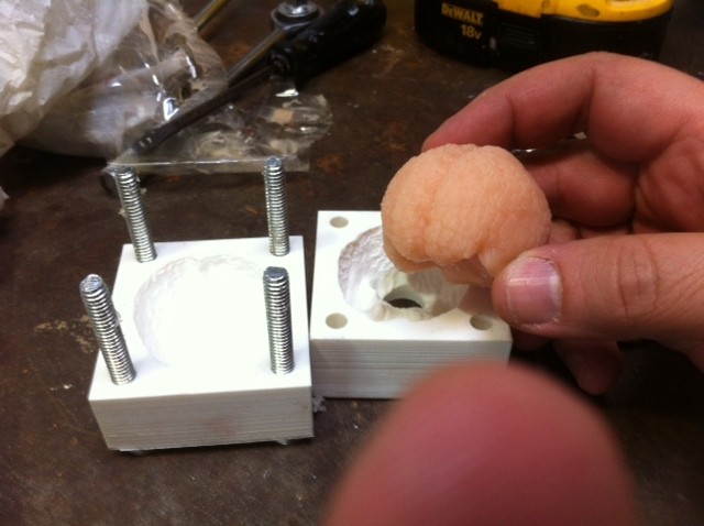
Professor Francis LeBouthillier involved me in this important and exiting project to help with seperating/analyzing organs and recreating the fetuses from CT Scans to printable 3D models
Abstract
Medical surgeons need to hone their skills prior to performing high-risk surgical procedures on fetuses in utero and on newborns. To date, highly representational 3-dimensional fetal models, that have life-like physical properties, do not exist. The goal of our research project was to create a series of accurate and responsive fetal models to help surgeons visualize the complex physiology of a newly developing fetus and their associated pathologies. These models provide surgeons with the visual and tactile information necessary to confidently implement highly complicated fetal surgical procedures.

The Surgical Training Fetus model could be used to address such fetal surgeries as:
Fetal Oral Teratoma
Fetal Bladder Obstructions
Aortic or Pulmonary Valvuloplasty - opening the aortic or pulmonary fetal heart valves to allow blood flow
Atrial Septostomy - opening the inter-atrial septum of the fetal heart to allow unrestricted blood flow between the atriums
Critical Aortic Stenosis – narrowing of the main outlet valve of the left ventricle.
Congenital Diaphragmatic Hernia - Balloon tracheal occlusion
Spina Bifida - Fetoscopic closure of the malformation